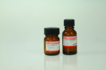
Efficient chemiluminescence kit
|
Cat # |
Product Name
|
Component
|
Pack Size
|
|
|
GE2301-25ML
|
Efficient chemiluminescence kit
|
Western HRP Substrate Luminol Reagent
|
12.5ml×2
|
250cm2
|
|
GE2301-50ML
|
25ml×2
|
500cm2
|
||
|
Western HRP Substrate Peroxide Solution
|
||||
|
GE2301-100ML
|
50ml×2
|
1000cm2
|
||
Introduction
Chemiluminescent detection uses an enzyme to catalyze a reaction that results in the generation of visible light. The horseradish peroxidase(HRP) chemluminescent reaction is based on the catalyzed oxidation of luminol by peroxide.Oxidized luminol emits light as it decays to its ground state. This technique has the speed and safety of chromogenic detection methods, at higher sensitivity levels.
Western HRP Substrate provides high sensivity in western or dot/slot/spot blotting application on both PVDF and nitrocellulose transfer membranes, and is compatible with all commonly used buffers and blocking reagents. Blots on PVDF membrane may be reprobed, allowing detection of multiple target proteins on the same blot.
The HRP substrate consists of Luminol Reagent and Peroxide Solution. Working HRP substrate is prepared by combining equal volumes of Luminol Reagent and Peroxide Solution. The HRP substrate produces a high intensity signal with low background for detection of both high and low background for detection of both high and low abundance proteins.
Materials Required for Western Blotting
1. PVDF membrane or nitrocellulose membrane.
2. Sheets of filter paper, cut to dimension of the gel.
3. 100% methanol (to pre-wet PVDF membrane).
4. Transfer system and transfer buffer.
5. ddH2O water.
6. Wash buffer: Phosphate-buffered saline(PBS) or Tris-buffered saline(TBS) containing 0.05-0.1% Tween-20 surfactant; (PBS:10 mM sodium phosphate, 150 mM NaCl, pH7.2. TBS: 10 mM Tris, 150 mM NaCl, pH7.4).
7. Blocking buffer:1-5%(w/v) blocking agent (e.g., casein, BSA, or nonfat dry milk ) in wash buffer.
NOTE: Western HRP Substrate is compatible with all blocking buffer.2% casein solution is recommended for the lowest background and highest signal-to-noise ratio.
8. Primary antibody specific for the protein of interest, diluted in wash buffer or blocking buffer.
9. HRP-conjugated secondary antibody, specific for primary antibody, diluted in wash buffer or blocking buffer.
10. Shallow trays, large enough to hold the blot.
11. Plastic wrap, plastic bag, transparency or sheet protector.
12. X-ray film and developer reagents or chemiluminescence-compatible imaging systems.
Usage Guidelines
1. Due to the high sensitivity of the Western HRP Substrate, lower amounts of antigen and higher dilutions of primary and secondary antibodies are recommended.Typical primary antibody dilutions are 1:1000-1:20000 and secondary antibody dilutions typically range from 1:20000-1:200000. Important: If switching to Western HRP Substrate from a lower sensitivity substrate, previous antibody dilution factors may need to be increased at least five-fold for the primary antibody and two-to five-fold for the secondary antibody to achieve the optimal signal-to-noise ratio.
2. Optimization of blocking reagents and incubation times will improve results and should be determined experimentally.
3. The high sensitivity of the Western HRP Substrate may result in a significant reduction in required x-ray film exposure time. An initial exposure time of 30 seconds is recommended. Optimum exposure time should be determined for each antibody system.
4. Always wear gloves and use blunt tip forceps when handling the membrane to avoid contamination.
5. Use care when handling the membrane to prevent tearing.
6. Do not use sodium azide, which inhibits HRP activity, in any buffer or reagents.
7. Use of blocking buffer to dilute antibodies may reduce background and increase sensitivity.
Western Blotting Protocol
Protein Transfer
1. Resolve the protein mixture on a 1-D or 2-D polyacrylamide gel.
2. Immerse the gel in an appropriate transfer buffer and allow it to equilibrate for 10-15 minutes.
3. If working with a PVDF membrane: Wet the membrane in 100% methanol for 15 seconds, or until the membrane appearance changes uniformly from opaque to semitransparent.
If working with a nitrocellulose membrane: Proceed to step 4. Nitrocellulose do not require prewetting.
4. Equilibrate the membrane for at least 5 minutes in the transfer buffer.
5. Soak filter paper in the transfer buffer for at least 30 seconds.
6. Assemble the transfer stack as shown below.
NOTE: To ensure an even transfer, remove air bubbles by carefully rolling a clean pipette over the surface of each layer in the stack. Avoid excessive pressure that can damage the gel and membrane.
7. Transfer proteins according to blotting apparatus manufacturer’s instructions.
8. Remove the blot from the transfer system and briefly rinse the membrane in ddH2O water. to remove gel debris. Proceed with immunodetection protocol below. If required, the PVDF membrane blot may be air dried and stored refrigerated for several months.
Antibody Incubations
1. If PVDF membrane were dried after transfer, wet the blots in 100% methanol for 15 seconds, or until the membrane appearance changes uniformly from opaque to semitransparent.
NOTE: Omit this step if using nitrocellulose membrane.
2. Rinse the blot with water and then place the blot in blocking buffer and incubate for 1 hour with gentle agitation at room temperature.
3. Prepare primary antibody solution by diluting the antibody in wash or blocking buffer. See Usage Guidelines for antibody dilutions.
4. Place the blot in the diluted primary antibody solution and incubate for at least 1 hour with gentle agitation. Ensure that the solution moves freely across the entire surface of the membrane.
5. Wash the blot with fresh wash buffer a minimum of three times with gentle agitation for 5-10 minutes. Additional or longer washes may further reduce background. Immobilon-p (0.2μm) membrane may require additional washing due to its greater surface area.
6. Prepare HRP-conjugated secondary antibody solution by diluting the antibody in wash or blocking buffer. See Usage Guidelines for antibody dilutions.
7. Place the blot in the diluted HRP-conjugated secondary antibody solution, and incubate for 1 hour with gentle agitation. Ensure that the solution moves freely across the entire surface of the membrane.
8. Wash the blot with fresh wash buffer a minimum of three times with gentle agitation for 5-10 minutes. Additional or longer washes may further reduce background.
Chemiluminscent detection
1. To prepare working HRP substrate, mix equal volumes of Luminol Reagent and Peroxide Solution in a clean container or test tube. Approximately 0.1 mL of working HRP substrate is required per cm2 membrane area.
The volumes of working HRP substrate needed for some common membrane sizes are indicated below:
|
Blot Size |
Working HRP Substrate Required |
|
7×8.5cm |
6ml (3ml Luminol reagent) (3ml peroxide solution) |
|
10×10cm |
10ml (5ml Luminol reagent) (5ml peroxide solution) |
|
8.5×13.5cm |
12ml (6ml Luminol reagent) (6ml peroxide solution) |
2. Allow the HRP substrate to reach room temperature (~10 minutes). Protection from light is not required.
3. Place the blot protein side up in a clean container, and add the HRP substrate onto the blot.
4. Incubate the blot for 5 minutes at room temperature.
5. Drain the excess substrate.
6. Cover the blot with a clean plastic wrap or sheet protector and remove any air bubbles. Ensure that the surface of the plastic wrap or sheet protector is dry and unwrinkled.
7. Expose the blot to a suitable X-ray film for an appropriate duration. Because of the high sensitivity of the Western HRP Substrate, a shorter exposure time may be required. The recommended initial exposure time is 30 seconds. The chemiluminescent,signal on the blot will last at least two hours. If necessary, fresh HRP substrate can be added to the same blot for consecutive exposures.
NOTE: The working HRP substrate can be stored up to 7 days in the dark at 2-8℃ without any detectable loss of activity.
Membrance Stripping (Only for PVDF Membrances)
A single blot on PVDF membrane can be sequentially probed by stripping the first antibody from the blot, and then incubating with a different primary antibody. This is especially useful for method optimization or when sample amount is limited.
The stripping process disrupts the antigen-antibody interaction,usually by a combination of detergent and heat or by exposure to low pH.
